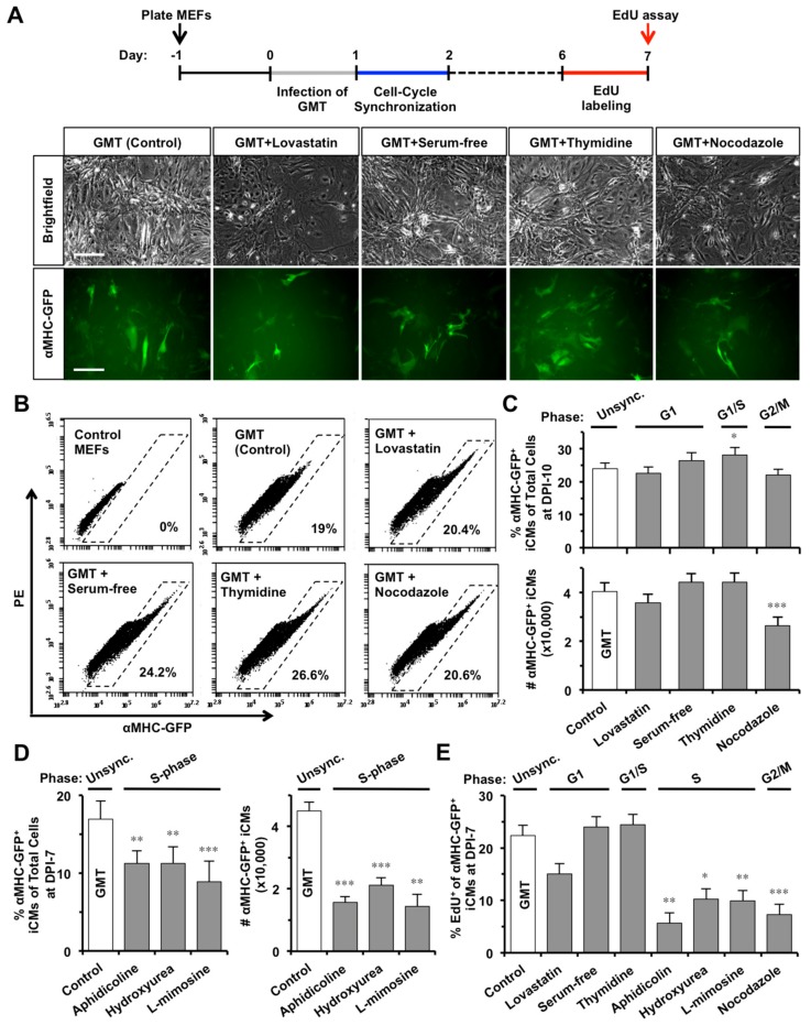
Figure 2.
S- or G2/M-phase synchronization at DPI-1 enhances cell-cycle exit in GMT-reprogrammed iCMs. (A) At DPI-1, MEFs were synchronized at G1, G0/G1, G1/S, or G2/M-phase by lovastatin, serum-free media, thymidine, or nocodazole, respectively. Representative pictures showing GMT-reprogrammed MEFs at DPI-10 with or without (Control) cell-cycle synchronization. Scale bars indicate 100 µm. (B) Representative FACS plots of reprogrammed αMHC-GFP+ iCMs at DPI-10. (C) The effect of G1-, G1/S-, or G2/M-phase synchronization on GMT-iCMs (n = 10), including the percentage (upper panel) and absolute number (lower panel) of αMHC-GFP+ iCMs at DPI-10. (D) The effect of S-phase synchronization by aphidicolin, hydroxyurea, or L-mimosine on GMT-iCMs (n = 5) at DPI-7. (E) The percentage of EdU+ cells in αMHC-GFP+ iCM-population at DPI-7 with or without cell-cycle synchronization at DPI-1 (n = 3). * p < 0.05, ** p < 0.01, *** p < 0.001 vs. GMT group.

