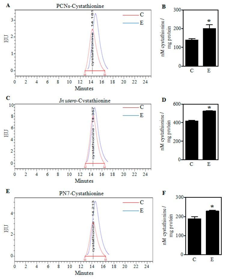
Figure 1.
Effect of ethanol on cystathionine levels in PCNs and fetal brain cortices. A representative high-pressure liquid chromatography (HPLC) profile of cystathionine in Control (C) and ethanol (E)-treated PCNs (A); The concentration of cystathionine quantified using standards in PCNs (n = 6) (B); Pregnant rats (Sprague-Dawley) at embryonic day 17 (ED 17) were administered five doses of E (3.5 g/kg b.wt.) or isocaloric dextrose (Control) by gastric intubation at 12 h intervals. At ED 19 the brain cortex from embryos was dissected, the protein samples were HPLC analyzed for cystathionine. A representative cystathionine HPLC chromatogram of fetal brain cortices obtained from Control and E-exposed pregnant rats (C); Cystathionine concentration quantified with appropriate standards of the HPLC profiles of panel C (n = 3) (D); HPLC-based determination of cystathionine in brain cortices of postnatal day 7 (PN7) ethanol pups that received a oral dose of 4 g/kg body weight of 20% v/v ethanol (in milk solution) split into 2 feedings at 2 h interval with the controls receiving iso-caloric and iso-volumic equivalent maltose-dextrin milk solution substituted for ethanol (E); Fetal brain cortex cystathionine content quantification measured by HPLC in the PN7 model (n = 3) (F). Values represent the mean ± SEM. * p < 0.05 was considered significant vs. ethanol.

