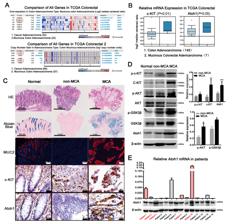
Figure 4.
Expressions of c-KIT and Atoh1 in CRC patients. (A) Compared with cecum adenocacinoma (n = 22), mRNA levels of MUC2 was markedly increased in mucinous colon adenocarcinoma (n = 22) in TCGA Colorectal (red boxes, p-value). DNA copies of c-KIT and Atoh1 were significantly increased in mucinous colon adenocarcinoma (n = 37) indicated by TCGA Colorectal 2; (B) the mRNA levels of c-KIT and Atoh1 were also elevated in MCA patients (n = 145) compared with non-MCA (n = 7) from TCGA Colorectal. p < 0.05 or 0.01; (C) according to the mucus area estimated under HE and Alcian blue staining, human CRC tissues were divided into non-MCA and MCA. Paratumoral normal tissues were used as controls. MUC2 was filled in goblet cells in normal colon and the main mucus component in MCA, while there was rare MUC2 in non-MCA tissues. Normally, c-KIT was expressed in the membrane while Atoh1 in the nuclei of epithelial cells, including goblet cells. Compared with non-MCA, c-KIT and Atoh1 were remarkably elevated in MCA, indicated by much more intensive immunostaining; (D) protein expressions of total c-KIT, p-c-KIT, and Atoh1 were clearly higher in MCA patients (n = 5) than those in non-MCA patients (n = 9) combined with an increased expression of p-AKT and p-GSK3β. * p < 0.05, ** p < 0.01, *** p < 0.001; and (E) real-time PCR was performed to detect Atoh1 mRNA level in CRC tissues from 14 patients, including 5 MCA patients marked by red color. Glyceraldehyde-3-phosphate dehydrogenase (GAPDH) was used as the internal control. Normalized Atoh1 mRNA expression was shown in the column graph. Western blot showed the protein expression of Atoh1 which was consistent with its mRNA level except patient 3. All values are mean ± SEM of three independent experiments unless otherwise stated.

