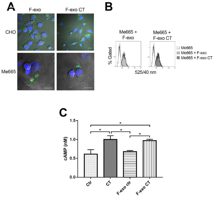
Figure 5.
F-exo transfer to cells induces increase of cellular cAMP. (A) Confocal fluorescence microscopy images of F-exo and F-exo CT transfer on CHO and Me665 cells. 2 × 10⁸ fluorescent exosomes were incubated with 4 × 10⁴ CHO and Me665 cells for 4 h at 37 °C. Cells were then fixed and analysed; Scale bars represent 20 µm; (B) FACS analysis of F-exo and F-exo CT transfer on target cells. 4 × 10⁴ cells were incubated with 1.5 × 10⁷ F-exo or F-exo CT for 4 h at 37 °C. At the end of the incubation cells were FACS analysed. The increase in cell fluorescence demonstrate exo transfer to cells; (C) cAMP assay of Me665 cells treated with F-exo. 2 × 10⁵ cells were incubated with 5.7 × 10⁷ F-exo, F-exo CT or 0.2 ng/mL CT for 4 h at 37 °C. The graph shows the intracellular cAMP production of cells. * p < 0.05 Values are means ± S.D. (n = 3).

