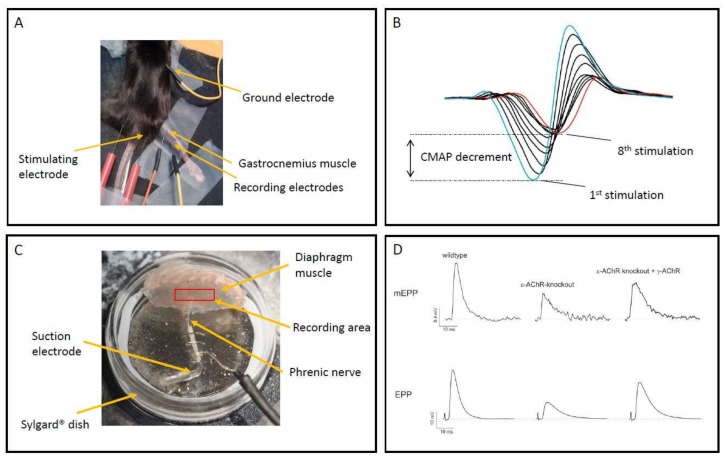
Figure 1.
Panel (A) shows experimental setup for in-vivo electromyography of anaesthetised mouse, with location of stimulating and recording mono-polar needle electrodes. Panel (B) shows example trace of compound muscle action potential (CMAP) recorded for gastrocnemius muscle, significant decrement is evident between 1st and 8th stimulation at 10 Hz. Panel (C) shows experimental setup for sharp electrode recording from ex-vivo mouse phrenic nerve/hemi-diaphragm muscle, central area surrounding phrenic nerve branch within muscle where recordings are acquired is indicated. Panel (D) shows examples of miniature endplate potentials (mEPPs) and stimulated endplate potentials (EPPs) recorded from an 8-week-old wildtype mouse, an ε-AChR knockout mouse and an ε-AChR knockout mouse with human γ-AChR knocked-in. ε-AChR knockout mice have severely reduced mEPP and EPP amplitude due to diminishing post-natal expression of γ-AChR containing receptors with no adult AChR expression, knock-in of human γ-AChR partially restores mEPP and EPP amplitude and is a model for AChR-deficiency CMS.

