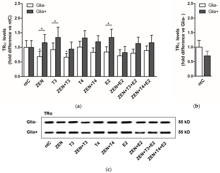
Figure 2.
TRα protein expression levels in cerebellar granule cells in the absence, Glia−, or presence of glial cells, Glia+; treated with zearalenone (ZEN), and/or triiodo-thyronine (T3), thyroxine (T4) and 17β-estradiol (E2) for 18 h, examined by Western blotting; (a) Shown P-values were calculated: compared to ntC (above and next to the bars) and Glia+ compared to Glia− (*) p < 0.05 (above braces) in each treatment group; (b) Expression of TRα protein in non-treated controls normalized to Glia− (p-value not shown). All data represents the mean ± SD of at least three independent experiments (n = 6 per treatment); (c) Representative Western blot images.

