Figure 4.
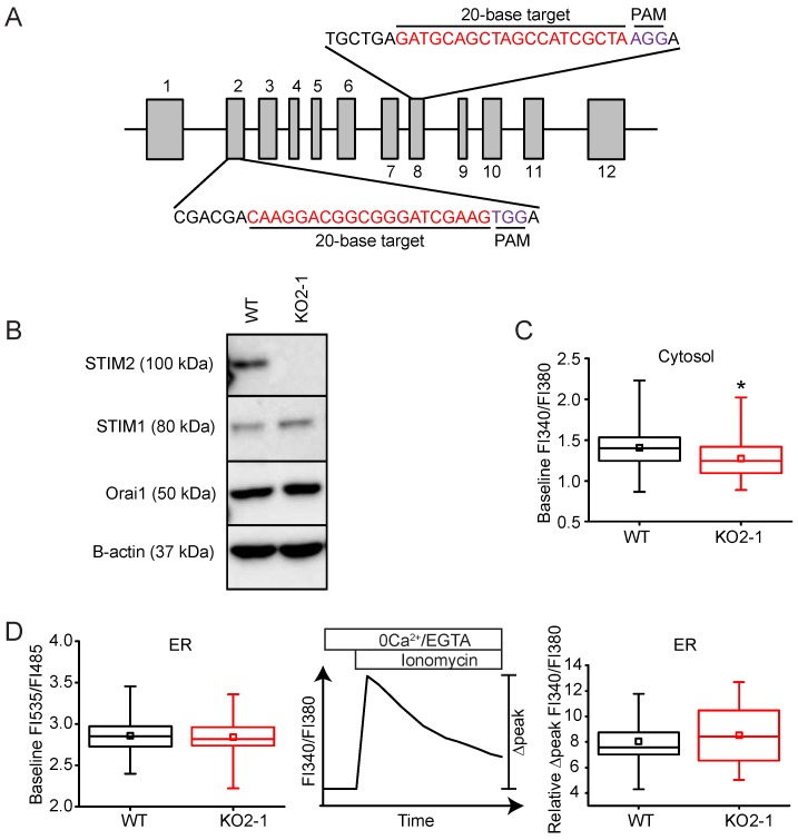
STIM2 knock-out in NIH 3T3 cells does not alter Ca2+ homeostasis. (A) Exon map of murine Stim2 showing the 20-base-pair gRNA CRISPR-Cas9 targets in exons 2 and 8 used in the studies. (B) Western immunoblot of whole cell protein lysates (20 μg) harvested from control (WT) or Stim2-null (KO2-1) NIH 3T3 cells. Lysates were resolved by 8% SDS-PAGE and identified using selective antibodies against STIM2, STIM1, Orai1, and β-actin. (C) Baseline FI340/FI380 in WT (n = 172 cells) and KO2-1 (n = 294 cells) NIH 3T3 fibroblasts. * p < 0.05 compared to WT (D) STIM2 KO did not alter basal [Ca2+]ER as measured by two independent methods. Left Panel: Comparison of [Ca2+]ER in WT (n = 125 cells) and KO2-1 (n = 101 cells) cells using D1ER, a genetically-encoded biosensor of ER Ca2+. Middle Panel: Response of [Ca2+]c following release of intracellular Ca2+ stores with ionomycin (2 µM). Right Panel: The increase in cytosolic Ca2+ (Relative ∆peak FI340/FI380) following ionomycin-induced discharge of ER Ca2+ in the Stim2 null cells was similar to that measured in control cells (KO2-1: n = 62 cells; WT: n = 61 cells).

