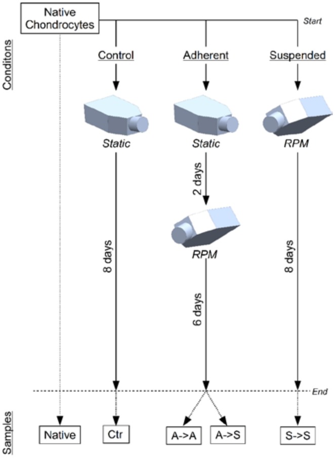
Figure 1.
Experiment design. Bovine chondrocytes were distributed to six experimental groups in commercial T25-flasks. The control group was left for eight days in static monolayer culture (Ctr). The “adherent” groups were kept for two days in monolayer culture and subsequently exposed to the RPM for six days. The “suspended” groups were immediately placed on the RPM for eight days (S->S). After the experiment, the samples were collected for further analysis. For the “adherent” groups, the adherent cells (A->A) were collected separately from the cells that became suspended (A->S). In addition, freshly thawed cells were lysed for gene expression analysis, which was considered to represent the “native” chondrocytes.

