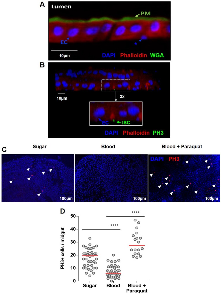
Fig 1. General structure of the midgut epithelium of Aedes aegypti and modulation of cell proliferation upon blood meal.
The midgut epithelia from a blood-fed A. aegypti females were fixed in PFA and in (A) sections of 0.14 μm were stained with WGA-FITC (green), red phalloidin (red) and DAPI (blue). The peritrophic matrix (PM), intestinal lumen (Lumen), polyploid enterocytes (EC) and basally localized–putative proliferative cells (*)–are visible. In (B), confocal image (z-stack of 0.7 μm slides (20X)) of the two monolayers of the midgut of a blood-fed female, 5 days post feeding, stained with PH3 mouse antibody (green), DAPI (blue), and phalloidin (red)–Inset (2x): polyploid enterocytes (EC) are PH3-positive ISC (ISC) are visible. (C) Mosquitoes were fed on a sugar solution (10% sucrose), blood or blood supplemented with 100μM of the pro-oxidant paraquat. The insect midguts were dissected 24 hours after feeding and immunostained for PH3. Representative images of mitotic (PH3-labeled) cells (red) in the epithelial midgut of animals fed on sugar, blood or blood supplemented with paraquat are shown. The nuclei are stained with DAPI (blue). The arrowheads indicate PH3+ cells. (D) Quantification of PH3-positive cells per midgut of sugar, blood or blood plus paraquat-fed mosquitoes for sugar and blood and 18 for blood-paraquat fed midguts. The experiments were performed on Red Eye mosquito strain. The medians of at least three independent experiments are shown (n = 40 for sugar and blood and n = 18 for paraquat supplemented blood). The asterisks indicate significantly different values, **** P<0.0001 (Student’s t-test).

