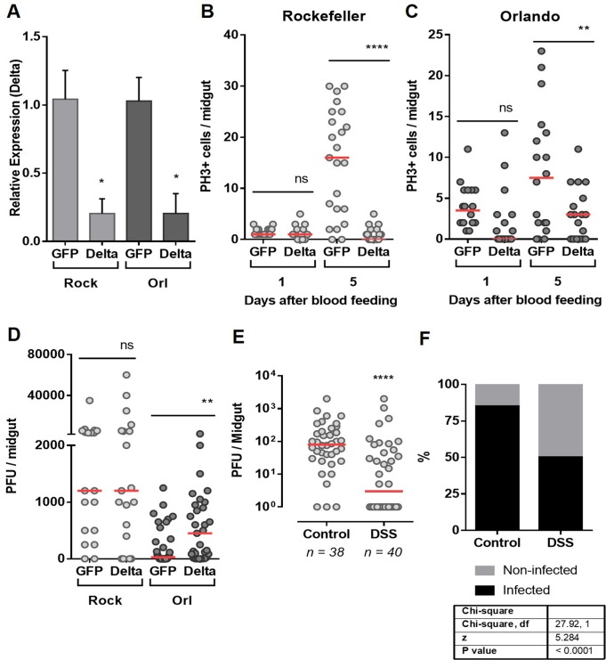
Fig 4. Interference in gut homeostatic response impacts vector competence.
(A). The midguts of dsRNA-injected Rockefeller and Orlando mosquitoes were dissected 24 days after a blood meal for silencing quantification of Delta, the ligand of Notch. Total PH3-positive cells were quantified from midguts of silenced Delta or control (GFP) mosquitoes from the Rockefeller (B) or Orlando (C) strains, both 1 and 5 days after blood meal. (D) dsRNA-Injected mosquitoes were fed DENV2-infected blood, and 5 days after the infection, the midguts were dissected for the plaque assay. (E) The susceptible (Rockefeller) mosquitoes were pre-treated with the tissue-damaging dextran sulfate sodium (DSS) accordingly to material and methods section. Twelve hours after the end of the DSS treatment, the mosquitoes were fed with DENV-2-infected blood. After 5 days, the midguts were dissected for the plaque assay. (F) The percentage of infected midguts (infection prevalence) was scored from the same set of data as in (E). The medians of at least three independent experiments are shown. n = 20–25 in (A), (B) and (C); n = 20–26 in (D) and n = 40 in (E). Statistical analyzes used were: Student’s t-test for (A), (B) and (C); Mann-Whitney U-tests were used for infection intensity (D and E); and chi-square tests were performed to determine the significance of infection prevalence analysis (F). *P<0.05, ** P<0.01, **** P<0.0001.

