Figure 1.
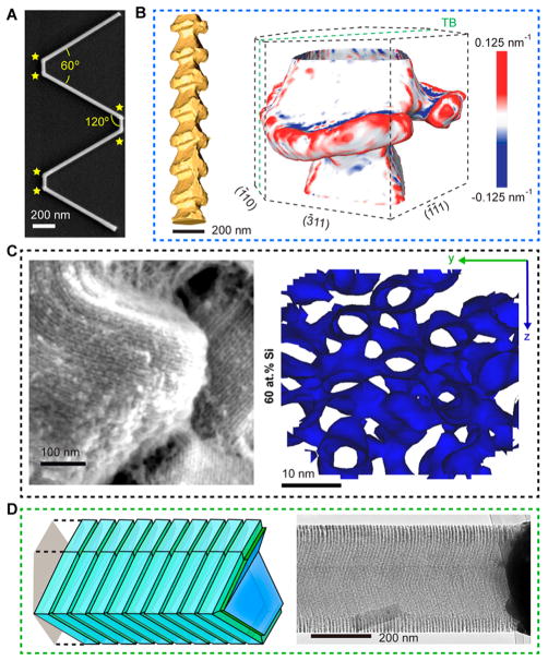
New Si nanostructures can be explored for enhancing subcellular biointerfaces. (A) SEM image of a multiply kinked SiNW, showing six kinks (yellow stars) with individual angles of 120°. (B) STEM tomography of a Si spicule, showing anisotropic features (left); 3-D curvature map of one segment (right) shows convex and concave features in the spicule. “TB” denotes twin plane. Adapted with permission from ref 13. Copyright 2015 American Association for the Advancement of Science. (C) SEM (left) and 3D atom probe tomography isosurface (right) images of mesoporous Si particles, revealing periodic arrangements of Si nanowire assembly (left) and 3D structures interconnected by microbridges (right). The isosurface is plotted at 60 wt % Si. Adapted with permission from ref 8. Copyright 2016 Macmillan Publishers Ltd. (D) A schematic diagram (left) and a TEM image (right) of a SiNW with massively parallel sidewall grooves. Adapted with permission from ref 32. Copyright 2017 The Authors.

