Figure 1.
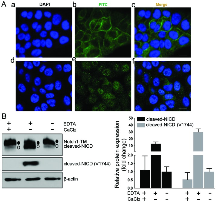
Validation of Notch1 antibody for detection of membranous and nuclear Notch1 in HN4 cells. (A) Immunofluorescent staining was observed after HN4 cells were treated with PBS (a-c) or 2.5 mM EDTA (d-f) for 10 min and immunostained with the anti-Notch1 antibody. The cells treated with PBS demonstrated strong membranous staining of Notch1 (a-c). An obvious nuclear enrichment of Notch1 staining was observed in the cells treated with EDTA (d-f). Scale bars, 10 µm. (B) Western blot analysis (left) and the quantification (right) of protein extracted from HN4 cells with different treatments revealed transmembranous (TM) Notch1 (S2 cleaved and S3 uncleaved, solid dot) and activated NICD (S3 cleaved, hollow circle). In PBS-treated HN4 cells, the Notch1 protein was mostly in the transmembranous form. The EDTA treatment induced NICD S3-cleaved, as a smaller size band was detected by the Notch1 antibody, and the cleaved state was further confirmed by the Notch1 Val1744-specific antibody. The treatment with CaCl2 neutralized the function of EDTA and reversed the S3-cleaved status induced by EDTA. The PBS-treated cells were set as the control group. The quantification analysis was calculated from three independent experiments.

