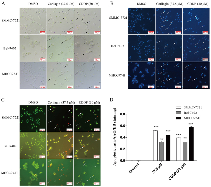
Figure 3.
Growth inhibitory effect of corilagin on hepatocellular carcinoma (HCC) cells. SMMC-7721 cells were treated with DMSO (0.1%, w/v) or 37.5 µM corilagin and CDDP as positive control for 24 h. (A) Representative images after treatments. Images were captured using phase-contrast microscopy (Leica DM IRB); arrows indicate apoptotic cells. Magnification, ×200. (B) Cells were fixed and stained using Hoechst 33258; apoptotic cells present brighter blue fluorescence; arrows indicate apoptotic cells. Magnification, ×200. (C) Cells were harvested and stained with AO/EB; arrows indicate apoptotic cells. Magnification, ×200. (D) The apoptotic ratio was determined in the AO/EB-stained apoptotic cells (**P<0.01 and ***P<0.001).

