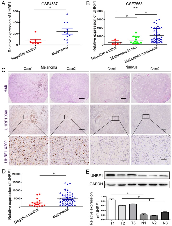
Figure 1.
UHRF1 is significantly upregulated in melanoma. (A and B) The expression of UHRF1 in the melanoma group is markedly upregulated compared with that of the control group by reexamining the GSE4587 and GSE7553 datasets. (C) Representative images of TMA stained with H&E and IHC for UHRF1. Scale bar, 50 µm. (D) The comparative expression of UHRF1 between the melanoma and negative control was analysed by IPP. (E) UHRF1 expression in melanoma (T1-T3) and benign nevi (N1-N3) was assessed by western blot analysis, and GAPDH expression was used as an internal reference (upper panel). Representative blots shown here were from experiments repeated three times with similar results (lower panel). *P<0.05; **P<0.01. T for tumor, N for normal tissues.

