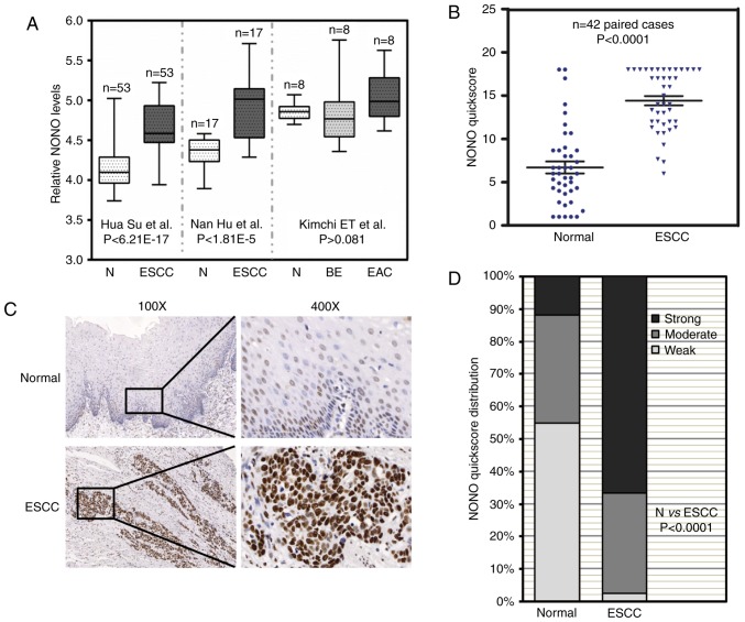
Figure 1.
Increased expression of NONO in ESCC tissue samples. (A) Three independent microarray data from the Oncomine database showed that NONO mRNA levels were increased in ESCC patient tissue samples. Comparison of NONO mRNA levels in normal tissue (N), esophageal squamous cell carcinoma (ESCC), and Barrett's esophagus (BE), esophageal adenocarcinoma (EAC) are presented together with P-values. (B) Immunohistochemistry signal of NONO from paired normal and ESCC tissue samples were recorded by quickscore method. Comparison of quickscore distribution between adjacent normal and ESCC samples was performed by χ2 test. (C) Representative immunohistochemistry (IHC) images of NONO in paired normal and ESCC tissue samples. NONO levels were lower in adjacent normal esophageal epithelium, but were higher in ESCC. (D) NONO quickscores were further divided into three groups: strong, moderate and weak. The percentage of each group in normal and ESCC tissue samples were plotted.

