
Figure 1.
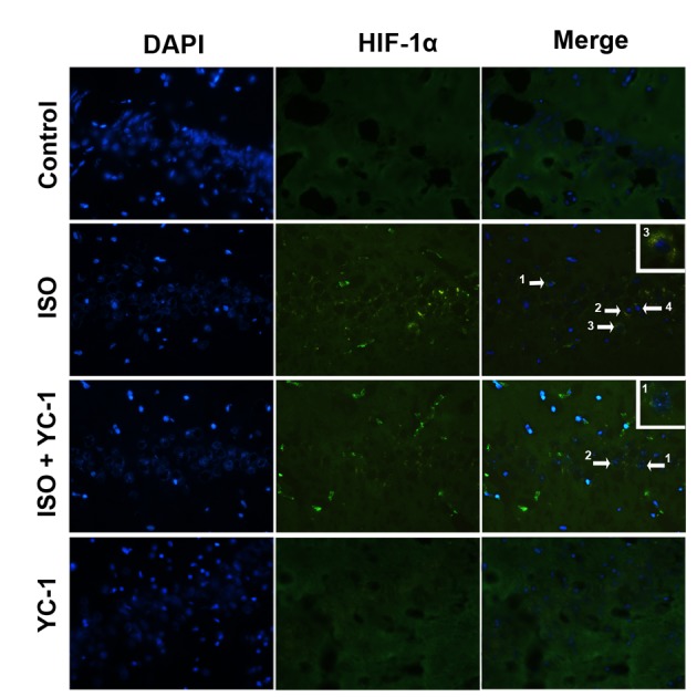
Immunofluorescence staining demonstrating distribution of HIF-1α (green) in the hippocampal CA1 area of aged rats. HIF-1α-positive cells in the hippocampal CA1 area were observed after 4-h isoflurane exposure. This staining was inhibited by YC-1, vs. the control group. n=6, Magnification, ×400. Scale bar=20 µm.