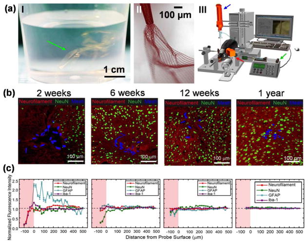
Figure 2.
Syringe delivery of mesh electronics into the brain to yield neuron interpenetration without a chronic immune response. (a) Unique structural and mechanical properties of mesh electronics allow for syringe delivery into the brain, highlighting a photograph of multiple mesh electronics probes (green arrow) floating in an aqueous saline solution similar to colloidal particles (I), a bright-field microscope image showing partially ejected mesh electronics with significant expansion in solution (II), and a schematic of controlled stereotaxic injection (III) that allows precisely targeted delivery of mesh electronics using a motorized translational stage for controlling needle withdrawal (blue arrow), a syringe pump for controlling the injection rate (green arrow), and a camera for visualizing the mesh during injection (red arrow) [14,36]. (b) Time-dependent immunohistochemical staining images of horizontal brain slices at 2 weeks (hippocampus), 6 weeks (cortex), 12 weeks (cortex) and 1 year (cortex) post injection. In all images of panel (b), red, green and blue colors correspond to neuron axons (Neurofilament antibody), neuron nuclei (NeuN antibody) and mesh elements. (c) Normalized fluorescence intensities plotted versus distance from the mesh/brain tissue interface at different time points; the intensities were normalized versus background far from the probe (black dashed horizontal lines). The pink shaded regions indicate the interior of mesh electronics [40].

