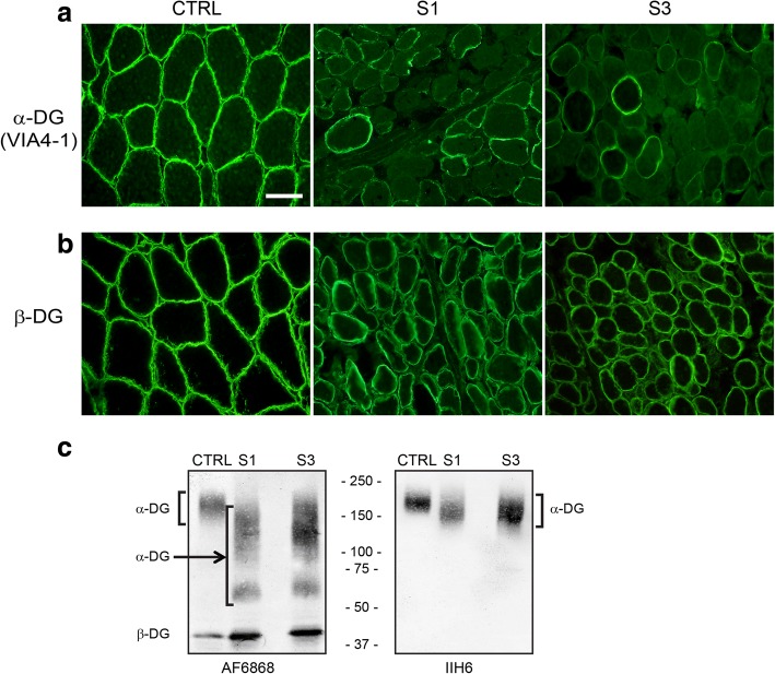
Fig. 2.
Subjects 1 and 2 display abnormalities in both α-dystroglycan staining and glycosylation. Control muscle or muscle taken from subject 1 (S1) and subject 3 (S3) were stained for alpha dystroglycan using VIA4-1 antibody (a) or β-DG (b). Note the reduced staining for α-DG but not β-DG in subjects 1 and 3. The size bar denotes 50 μm for all panels in a and b. c Western blot analysis of muscle tissue from control and subjects 1 and 3. Samples were probed with peptide-specific antibody AF6868 and the glycoepitope-specific antibody IIH6 as indicated. The location of α-DG and β-DG is indicated. Note that control shows a higher molecular size immunoreactive species for α-DG with both antibodies while S1 and S3 show a more heterogeneous species of much smaller molecular size, suggesting hypoglycosylation of the protein

