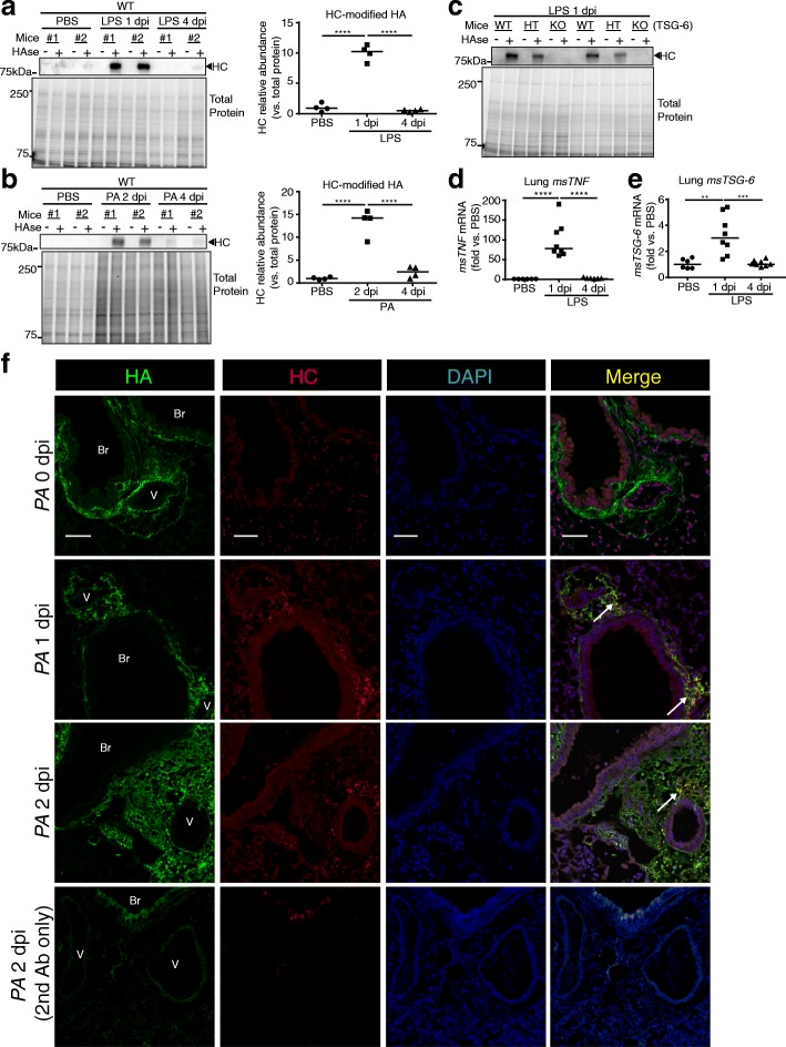
Fig. 1.
HC-HA formation after LPS or PA injury. a-c. Abundance of heavy chain (HC)-linked HA in lung lysates detected by western blot using IαI antibody (recognizing HC) on lungs before (−) and after (+) hyaluronidase (HAse), which releases HC linked to HA. Each lane represents an individual mouse lung exposed to intratracheally instilled LPS (20 μg; a) or Pseudomonas aeruginosa (PA, 2*106 CFU; b) or control PBS for the indicated time, noted as days post instillation (dpi). Lung HC abundance was expressed relative to that of total protein, measured by densitometry (a-b). c. Exclusive role of TSG-6 in forming HC-HA was confirmed using wild type (WT), heterozygous (HT), and knockout (KO) for TSG-6. d-e. msTNFα and msTSG-6 expression levels measured by qPCR in whole lung following LPS. Data in a-b and d-e analyzed by ANOVA with Tukey’s multiple comparisons; **P < 0.01, ***P < 0.001, ****P < .0001. f. Immunofluorescence images of HA and HC localization in formalin-fixed, paraffin-embedded lung sections from control (0 dpi) and PA-injured (1 and 2 dpi) mice, using antibodies against HA binding protein (red) or HC2 (green), and staining for nuclei with DAPI (Blue). Staining control provided in the last row, using secondary antibody only. Note HC-HA co-localization in the peri-broncho (Br)-vascular (V) interstitium (white arrow); scale bar 50 μm.

