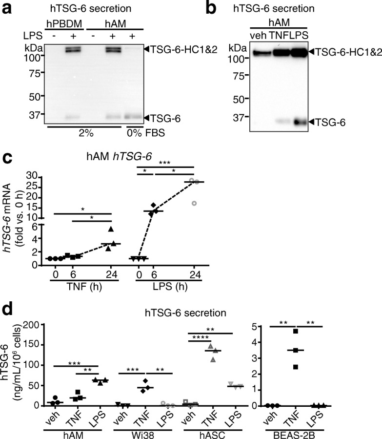
Fig. 2.

TSG-6 induction by TNFα or LPS stimulation of lung cells. a. Presence of TSG-6 in conditioned media of cultured human peripheral blood mononuclear cell-derived macrophages (hPBDM) and in human alveolar macrophages (hAM) following LPS stimulation (50 ng/mL, 24 h) detected by western blotting with TSG-6 antibody. Note that TSG-6 forms covalent TSG-6-HC intermediates only in the presence of 2% FBS (which contains serum inter-alpha-inhibitor that provides HC1 and HC2). b-c. TSG-6 secreted protein in supernatants (b; 2% FBS) and mRNA expression (c) of hAM stimulated with TNFα (20 ng/mL, 24 h or indicated time) or LPS (50 ng/mL, 24 h or indicated time), or vehicle (veh) assessed by western blot and qPCR, respectively. d. Levels of TSG-6 protein secreted in supernatant of hAM, human lung fibroblasts Wi38, human adipose stromal/progenitor cells (hASC), and human bronchoepithelial cells BEAS-2B stimulated with TNFα or LPS, measured by ELISA. In c-d, each data point represents an independent experiment; data analyzed with ANOVA and Tukey’s multiple comparisons. *P < 0.05, **P < 0.01, ***P < 0.001, ****P < .0001
