Fig. 1.
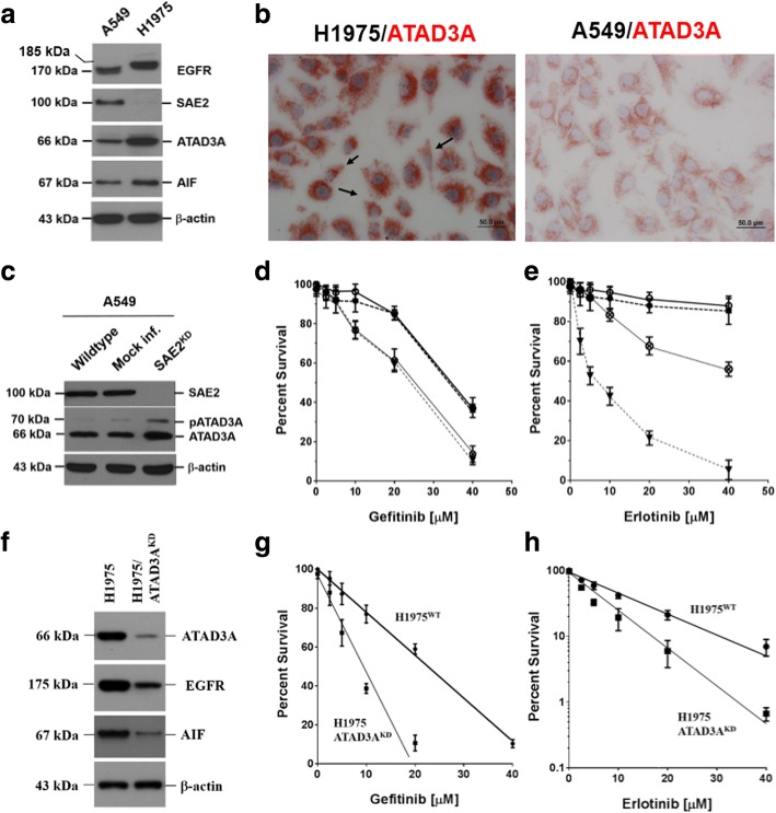
Expression of intracellular markers, SAE2 and ATAD3A, affect sensitivity of EGFR-mutated lung adenocarcinoma (LADC) cells to tyrosine kinase inhibitors, gefitinib or erlotinib. a LADC cells, carrying mutated EGFR (H1975), expressed abundant ATAD3A, an anti-apoptotic factor [9], as determined by a Western blotting analysis. b ATAD3A protein was highly expressed in EGFR-mutated H1975 (the left panel) and, intermediately, in the wild-type A549 (the right panel) cells. Some of the EGFR-mutated H1975 cells were elongated, scattered, and crescent shape (dark arrows), resembling phenotypes of mesenchymal cells [21], implicating that mutated EGFR could be associated with epithelial-to-mesenchymal transition (EMT) of LADC cells. c Knockdown of SAE2 expression (SAE2 ) increased ATAD3A expression. d In SAE2KD cells, gefitinib sensitivity was also increased, suggesting that SAE2 could be an intracellular sensor of gefitinib. It is therefore worth noting that gefitinib sensitivity of SAE2KD A549 cells was equivalent to that of H1975 cells (ID50 ~ 25 μM). e Silencing of SAE2 expression increased erlotinib sensitivity of A549 cells as well. However, SAE2KD A549 cells (ID ~ 50 μM) were more resistant than H1975 cells (ID ~ 5.6 μM) to erlotinib. f Silencing of ATAD3A (ATAD3A ) reduced expression of EGFR and AIF. g Increase of gefitinib sensitivity was thus anticipated in ATAD3AKD cells. h Increment of erlotinib sensitivity was also noted in ATAD3AKD cells

