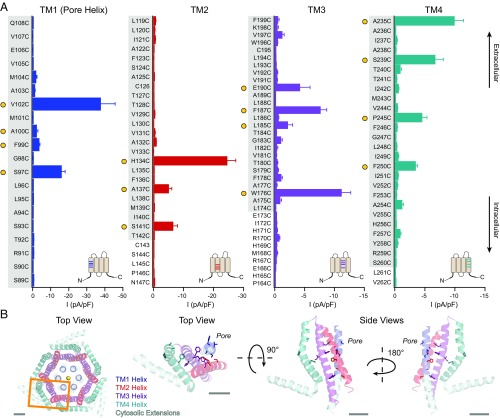
Fig. 1.
A cysteine screen of Orai1 TMs reveals several constitutively active mutants in all four TMs. (A) Current densities of Orai1 cysteine mutants in the absence of STIM1. For comparison, the current densities of WT Orai1 without and with STIM1 are −0.2 ± 0.01 pA/pF and −48 ± 8 pA/pF, respectively. Constitutively active mutants (defined as >2 pA/pF) are marked with filled yellow circles. Gray shaded areas on the labels indicate the boundaries of the membrane as represented in the dOrai crystal structure (7). (Insets) Approximate positions of the mutations on a topology diagram of Orai1 (n = 4–16 cells; values are mean ± SEM). (B) Top-down view of the crystal structure of dOrai with one subunit outlined in an orange box. A top-down view and two side views of one dOrai subunit are also shown. TMs 1–4 are colored in blue, red, purple, and teal, respectively, with the positions of constitutively open cysteine mutants represented as sticks. (Scale bars: 10 Å.)

