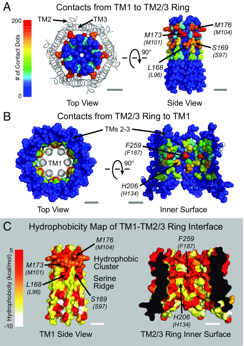
Fig. 6.
Atomic packing analysis and hydrophobicity mapping of dOrai reveals two distinct interfaces between the TM2/3 and TM1 segments. (A and B) Top and side view space-filling representations of the interface between TM1 and the TM 2/3 ring colored by the number of contacts per residue. TM4 is hidden for clarity. In A, TMs 2 and 3 are shown as ribbons. (C) Surface representation of the non–pore-lining residues of TM1 and residues of the TM2/3 ring facing TM1 colored according to amino acid hydrophobicity (47). A cluster of hydrophobic amino acids towards the extracellular side and a “serine ridge” towards the cytosolic side are also labeled. (Scale bars: 10 Å.)

