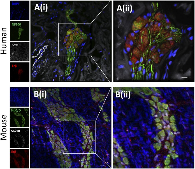
Fig. 1.
Erβ localization in mouse and human myenteric plexus. Two-dimensional projection of a z stack of confocal microscopy images of myenteric plexus from human (A) and mouse colon (B). (A) Human colonic myenteric ganglia stained for Erβ (red), Sox10 (gray), neurofilament (NF 200; green), and nuclei (DAPI; blue). Inset Ai is magnified in Aii. (B) Murine muscularis externa whole mount stained for Erβ (red), Sox10 (gray), HuC/D (green), and nuclei (DAPI; blue). Inset Bi is magnified in Bii.

