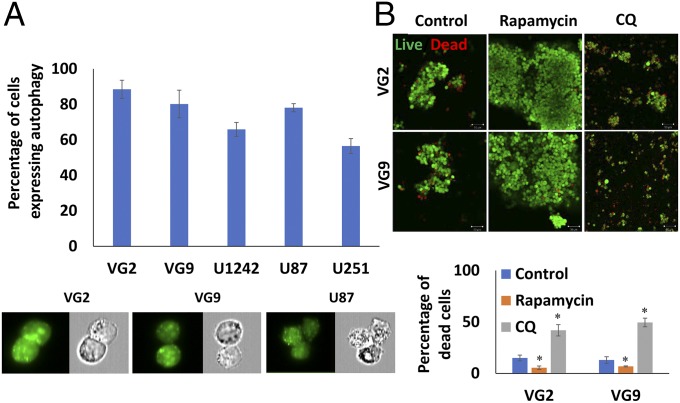
Fig. 1.
Autophagy is essential for survival of GSCs under anoikis conditions. (A) Percentage of autophagy in anoikis-resistant GSCs determined by flow cytometry. Fluorescence and phase-contrast images captured of the same cells [autophagic vacuoles (green) and blebbing/punctae]. (Magnification: 60×.) (B) Live/dead assay of neurospheres and GSC viability under conditions of autophagy induction (rapamycin treatment) and inhibition (CQ treatment) (green cells are viable). (Magnification: 100×.) Confocal images quantified for cell death and graphically represented. Error bars indicate ±SD, *P < 0.05.

