Fig. 3.
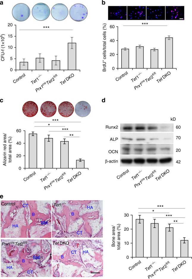
Tet DKO mice show BMMSC impairment. a Toluidine blue staining showed the CFU-F in control, Tet1−/−, Prx1creTet2fl/fl, and Tet DKO BMMSCs. b BrdU-labeling assay showed the proliferation rate in control, Tet1−/−, Prx1creTet2fl/fl, and Tet DKO BMMSCs. c, d When cultured under osteogenic inductive conditions, the capacities to form mineralized nodules of control, Tet1−/−, Prx1creTet2fl/fl, and Tet DKO BMMSCs were assessed by alizarin red staining (c), and the expression of osteogenic markers Runx2, ALP and OCN, as were assessed by western blotting (d). e New bone (B) formation of control, Tet1−/−, Prx1creTet2fl/fl, and Tet DKO BMMSCs when subcutaneously implanted into immunocompromised mice with hydroxyapatite tricalcium phosphate (HA/TCP; HA) as a carrier. *p < 0.05, **p < 0.01, ***p < 0.001 (mean ± SD). Scale bar, 50 μm. Results are from three independent experiments. p values were calculated using one-way ANOVA

