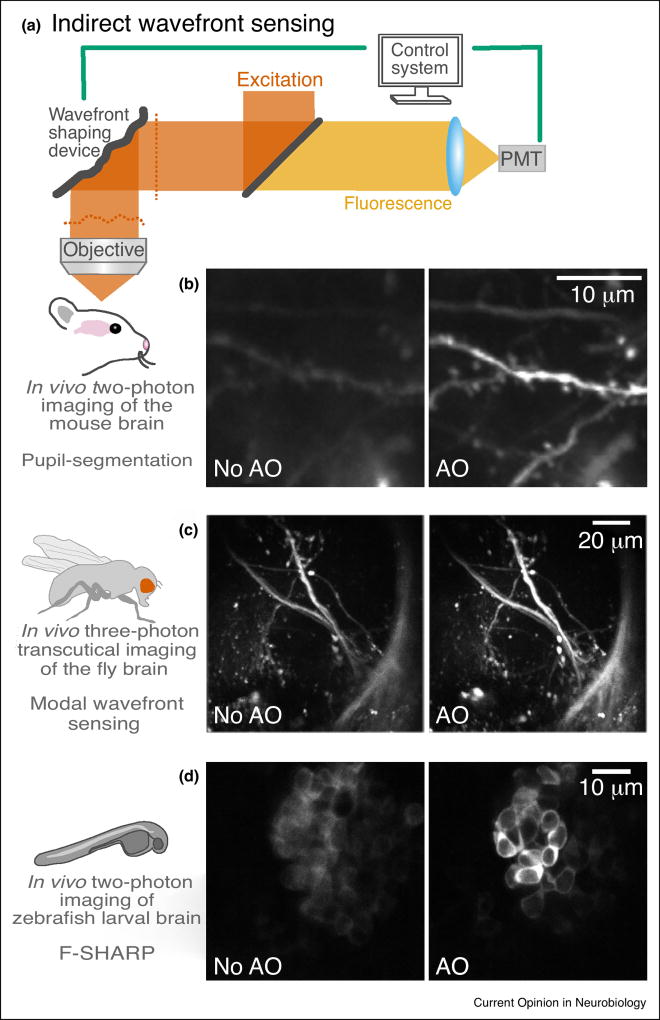
Figure 3.
Adaptive optical microscopy using indirect wavefront sensing. (a) An AO two-photon fluorescence microscope. PMT, photomultiplier tube. (b) Two-photon maximal-intensity projection images of dendrites at 376–395 µm below dura measured without and with AO in the mouse brain in vivo, using the frequency-multiplexed pupil-segmentation method [28••]. (c) In vivo three-photon transcutical imaging in the lateral horn of the fly brain [29•]. (d) Two-photon in vivo imaging of zebrafish larval brain obtained without and with AO correction, 300 µm under the brain surface, using F-SHARP [30••].

