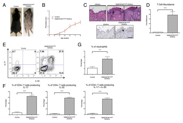
Figure 1. R26STAT3Cstopfl/fl CD4Cre mice developed a psoriatic-like skin phenotype.
(A) Representative image of 8-week-old R26STAT3Cstopfl/fl CD4Cre (skin score 3) and littermate R26STAT3Cstopfl/fl control mice. (B) Average skin phenotype scores of R26STAT3Cstopfl/fl CD4Cre (n=61) and control (n=20) mice from 2 to 8 weeks of age. The scale ranges from 0 (no phenotype) to 3 (greater than 50% fur loss and/or visibly dry/crusty skin). See methods for detailed description of the scoring system. (C) Representative histological skin sections from 8–week-old control and R26STAT3Cstopfl/flCD4Cre mice. Scale bar = 100 μm. Top middle panel: H&E staining. Arrow points to acanthosis and arrowhead points to parakeratosis in an area of hyperkeratosis. Top right panel: H&E staining. Arrow indicates an area reminiscent of Munro’s abscess. Bottom: Anti-CD3 staining, arrow highlights T cells tracking the epidermal/dermal boundary. (D) Fold difference of CD3+CD4+ T cells isolated from skin of R26STAT3Cstopfl/fl CD4Cre and control mice. (E) Representative intracellular flow cytometry analysis from CD3+CD4+ T cells isolated from the skin of 6–10 weeks old R26STAT3Cstopfl/fl CD4Cre and control mice. (F) Percentage of CD3+CD4+ skin T cells expressing IL-17 (left), IL-22 (middle), or both (right). (G) Percentage of neutrophils in the skin. For D–G, data is representative of 15 independent experiments with n ≥17 for each genotype, aged 6–10 weeks old. Statistical significance was assessed using (D) the Sign test and (F–G) the Mann-Whitney U test. Significance values are as follows: *** p ≤ 0.001; **** p ≤ 0.0001.

