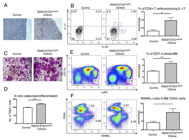
Figure 3. Th17-driven osteopenia phenotype of R26STAT3Cstopfl/fl CD4Cre mice was correlated with an increase in osteoclast progenitor cells and RANKL+ cells in the local environment.
(A) Representative immunohistochemistry staining of CD3 on the femur of control and R26STAT3Cstopfl/fl CD4Cre mice. (B) Representative intracellular staining of IL-17 and IL-22 in CD3+ CD4+ T cells in the bones of control and R26STAT3Cstopfl/fl CD4Cre mice (left) and quantified percentage of IL-17 producers in CD3+ CD4+ T cells (right). (C) Representative images of TRAP stained osteoclasts following in vitro osteoclast differentiation of bone marrow cells from R26STAT3Cstopfl/fl CD4Cre mice and their littermate controls. (D) Number of TRAP+ cells at day 10 of an in vitro osteoclast differentiation protocol normalized to number of TRAP+ cells derived from control bone marrow. (E–F) Flow cytometric analysis of OCP population and RANKL producers in the bone marrow of R26STAT3Cstopfl/fl CD4Cre mice and their littermate controls. All analysis was performed on mice aged between 6 to 10 weeks. Number of independent experiments ≥ 3 for all assays. Statistical significance was assessed using the nonparametric two-tailed Mann-Whitney U test. Significance values are as follows: ns p > 0.05; ** p ≤ 0.01; *** p ≤ 0.001; **** p ≤ 0.0001.

