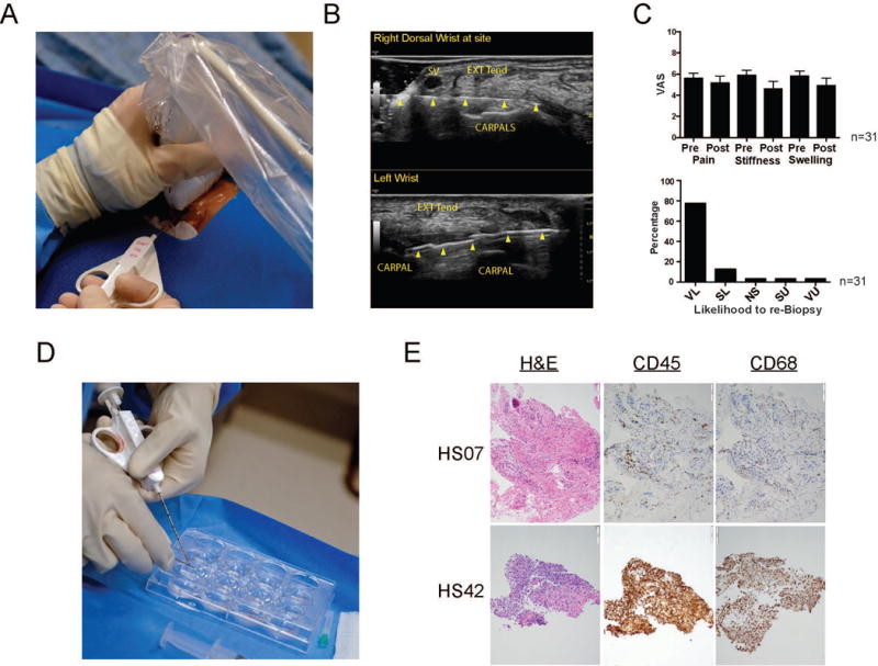
Figure 1.

Acquisition of synovial tissue from patients with RA. A. Ultrasound guided synovial biopsy from inflamed wrist with an 18-gauge by 1.5-inch needle. B. Dorsal transverse (axial) view of a right wrist (top) with a 25-gauge by 1.5-inch needle (arrowheads) and left wrist with an 18-gauge by 9 cm needle biopsy device with a 10-mm throw opened (2nd and 3rd arrows from left). SV = superficial vein, EXT TEN = extensor tendon complex, CARPALS = proximal row of carpal bones. C. Patients were asked prior to and following the procedure to complete a visual analogue score (VAS) assessing their pain, stiffness and swelling on a scale of 1-10. Post procedure patients were also asked their likelihood to agree to a subsequent procedure: (VL) very likely, (SL) somewhat likely, (NS) not sure, (SU) somewhat unlikely, and (VU) very unlikely. Error bars display SEM. D. Synovial tissue is removed from biopsy device and placed into PBS on ice until processed. E. Histomorphological features of synovial biopsy obtained from two representative RA patients. Representative photomicrographs of sections stained with Hematoxylin and Eosin (H&E), anti-CD45 (hematopoietic cells), and anti-CD68 antibodies (macrophages).
