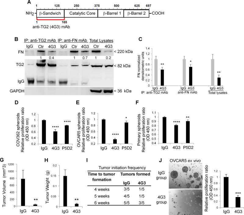
Figure 3. TG2/FN/Integrin β1 complex regulates spheroids proliferation and tumor initiating capacity.
A. Graphical representation of the epitope targeted by the 4G3 mAb overlapping with the FN-binding domain of TG2 (amino acids 1-165). B. Co-IP with anti-TG2 and anti-FN mAbs of cell lysates from OVCAR5 spheroids treated with 4G3 (10µg/ml) for 6 days. Western blotting was performed by using anti-TG2 and FN monoclonal antibodies. C. Densitometric analysis results are shown as means ± SEM. (N= 3; *P<0.05, **P<0.01). D–F. CCK-8 assay quantifies proliferation of spheroids derived from OC cell lines and primary cells treated with inhibitory mAbs directed against the FN-binding domain of TG2 (4G3), and integrin β1 (clone P5D2) (N= 8; *P< 0.05, **P< 0.01, ****P< 0.0001). G–H. Tumor weights and volumes derived from ALDH+/CD133+ sorted from OVCAR5 cells and treated with 4G3 or IgG control and injected sq in nude mice, as described (N= 5; **P< 0.01). I. Time to tumor formation for 10,000 ALDH+/CD133+ cells pre-treated with IgG or 4G3, grown as spheroids for 6 days, and injected sq in nude mice. J. Spheroid morphology (left panel) and proliferation assay (right panel) of cells isolated from xenografts and grown ex vivo. (N= 3; ***P< 0.001).

