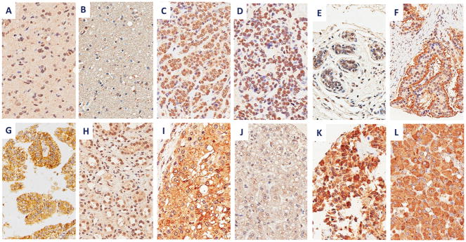
Figure 4.
Survivin immunohistochemistry with 2C2 mAb in human cancers and normal tissues with indicated staining patterns, including: (A) glioblastoma (nuclear), (B) normal brain (negative), (C) invasive ductal breast carcinoma (nuclear), (D) lobular breast carcinoma (nuclear), (E) normal breast tissue (negative), (F) typical carcinoid tumor (apical), (G) clear cell renal cell carcinoma (membranous and cytoplasmic), (H) normal kidney (nuclear), (I) hepatocellular carcinoma (membranous and cytoplasmic), (J) normal liver (negative), (K) medullary thyroid carcinoma (cytoplasmic), and (L) pheochromocytoma (cytoplasmic). Images are shown at 400x.

