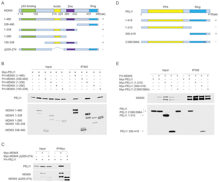
Figure 2. Mapping of interaction regions between Mdmx and Peli1.
(A) A schematic representation of Mdmx and the corresponding mutants (+: binding positive to Peli1; -: binding negative to Peli1).
(B) FH-Mdmx and the corresponding deletion mutants were individually transfected with Myc-Peli1 in H1299 cells. Whole cell lysates and immunoprecipitates with FLAG/M2 beads were subjected to western blot with anti-Myc (top) and anti-HA (lower) monoclonal antibodies.
(C) Myc-Mdmx and the Myc-Mdmx Δ200–274 mutant were individually transfected with FH-Peli1 in H1299 cells. Whole cell lysates and immunoprecipitates with anti-Myc antibody were subjected to western blot with anti-HA (top) and anti-Myc (lower) monoclonal antibodies.
(D) A schematic representation of Peli1 and the corresponding mutants (+: binding positive to Mdmx; -: binding negative to Mdmx).
(E) Myc-Peli1 and the corresponding mutants were individually transfected with FH-Mdmx in H1299 cells. Whole cell lysates and immunoprecipitates with FLAG/M2 beads were subjected to western blot with anti-HA (top) and anti-Myc (lower) monoclonal antibodies.

