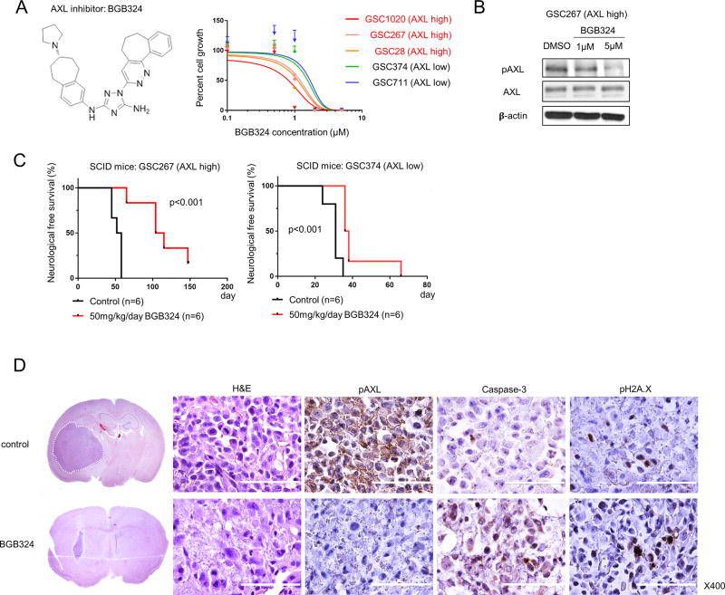
Figure 2.
BGB324 treatment inhibits AXL activity more effectively in cells with high levels of AXL expression. A. The molecular structure of BGB324, an AXL inhibitor (left) and a graphical representation showing that BGB324 inhibition of AXL in several GSCs results in a decrease in cell growth (right). The IC50 for BGB324 in cells expressing high levels of AXL is lower than that in cells expressing low levels of AXL. B. Western blot analysis shows that after 3 days of treatment with 1 or 5 µM BGB324, AXL is dephosphorylated in GSC267 cells. C. Kaplan-Meier survival curves generated from data obtained from xenograft mice inoculated with either GSC267 (AXL high) or GSC374 (AXL low) cells, with and without BGB324 treatment for 10 days (log-rank test). D. Representative hematoxylin and eosin (H&E) staining and IHC of GSC267 xenografts after 10 days of BGB324 treatment. In the H&E-stained coronal sections, white broken lines show the tumor area. Positive staining for Caspase 3 and pH2A.X indicates apoptosis. Scale bars, 100 µm.

