Figure 4.
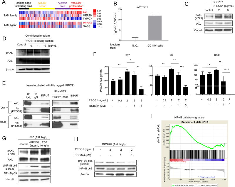
PROS1 activates AXL in GSCs. A. Heat map obtained from the IVY Atlas Project database showing the localization differences between TAM receptors and ligands. B. ELISA of mouse PROS1 (mPROS1) expression in conditioned medium from tumor-associated MG/Mø or medium alone (no cells, “N.C.”). Ten thousand cells, enriched for CD11b expression (as in Fig. 3D), were cultured in 1 mL RPMI-1640 medium for 48 hours, and the supernatant was used for the ELISA. C. Western blot analysis of total and phospho-AXL in GSC267 cells treated with 2 or 8 ng/mL of recombinant PROS1. D. Western blot analysis of total- and pAXL in GSC267 cells starved for 24h, followed by incubation with conditioned medium treated with 1 or 10 µg/mL of PROS1 blocking peptide. E. Immunoprecipitation (IP) analysis of GSC lysates using an AXL antibody (left panel) or Ni-NTA for PROS1 (right panel), followed by immunoblot for PROS1 and AXL, showing the AXL-PROS1 interaction. Cells were harvested 24 h after EGF, bFGF, and B27 starvation; lysates were then incubated with His-tagged rPROS1. Whole cell lysates with input rPROS1 served as a positive control, and pull-downs with immunoglobin G served as a negative control. F. Cell growth assays for three different GSCs (28, 267, and 1020) expressing high AXL levels treated with PROS1 and BGB324 for 24 h (* P < 0.05, ** P < 0.01, or *** P < 0.001). The cells were cultured in medium without EGF/bFGF and B27. G. Western blot analysis of total AXL, pAXL, and their downstream target, pNF-κB p65. Cells were starved of EGF/FGF and B27 for 24 h, then harvested after 30 min of treatment with 2 ng/mL PROS1 or 40 ng/mL EGF. Vinculin served as a loading control. H. Western blot analysis of pNF-κB p65 and total NF-κB after treatment with PROS1 with or without BGB324; cells were treated as above in (F). β-Actin served as a loading control. I. GSEA showing NF-κB signature is reduced in AXL-depleted GSCs. NES is shown in the plot.

