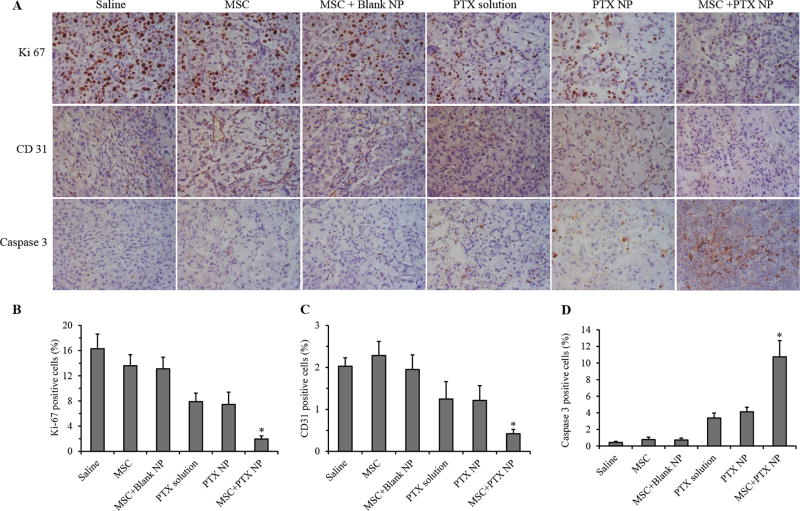
Figure 5. Immunohistological analysis of tumor sections.
(A) Tumors were collected at the end of the efficacy study and stained for Ki-67 (for tumor proliferation), CD31 (for angiogenesis), and caspase-3 (for apoptosis). Quantification of (B) Ki67, (C) CD31, and (D) cleaved caspase 3 staining. Data represented as mean ± SD, n = 6 images. * P < 0.01 compared to all other treatment groups.

