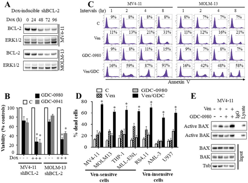
Figure 1. Dual inhibition of BCL-2 and PI3K pathway potently induced apoptosis in AML cells in association with a pronounced change in BAX conformation.

A) western blot analysis in MV4-11 and MOLM-13 cells displaying inducible knock-down of BCL-2 following exposure to 1 μg/ml doxycycline (Dox). B) These cells were left untreated or treated with doxycycline for 48 hr, and then exposed to the PI3K inhibitors GDC-0980 (500 nM) or GDC-0941 (1 μM) for 24 hr after which cell growth and viability was assessed by a CellTiter-Glo luminescent assay. Error Bars: S.D of 3 independent experiments; *, P < 0.05 in each case for values obtained in the presence vs absence of doxycycline. C) MV4-11 and MOLM-13 cells were exposed to venetoclax (10 nM) and GDC-0980 (500 nM) alone or together for the designated intervals after which the extent of apoptosis was determined by Annexin V staining. D) Various AML cell lines were exposed to ABT-199 and/or GDC-0980 for 24 hr after which the extent of cell death was determined employing Annexin V/PI staining. The agent concentrations used were: ABT-199: 2.5 nM for RS4,11; 10 nM for MV4-11 and MOLM-13; 50 nM for MLL-ENL; 100 nM for THP-1; and 500 nM for U937 and OCI-AML3 cells. GDC-0980: 1000 nM for U937 and RS4,11 cells; 500 nM for all other cell lines. Error Bars, S.D for at least 3 independent experiments; P < 0.003 for combined treatment compared to either agent alone for each cells. E) MV4-11 cells were exposed to venetoclax and/or GDC-0980 for 2 hr after which BAX and BAK conformational changes were assessed (top panel). Western blot analysis was also performed on input lysates (bottom panel).
