
Figure 6. Role of BAX, BAK, and BIM in venetoclax/GDC-0980 lethality in venetoclax-insensitive U937 cells.
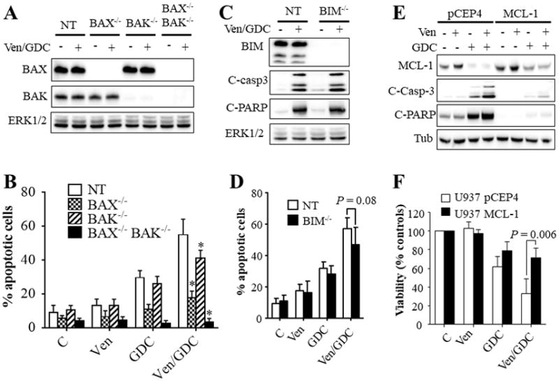
Cells exhibiting knockout of BAX, BAK, double BAX/BAK (A–B), or BIM (C–D), or non-targeting (NT) control cells were exposed to venetoclax (500 nM) and/or GDC-0980 (1.5 μM) for 16 hr. The cells were then lysed, and the lysates subjected to western blot analysis (A, C). Alternatively, the extent of apoptosis was determined using the Annexin V staining (B, D). Error Bars, S.D of at least 3 independent experiments. For B: P = 0.0005; P = 0.0223; P < 0.0001 for BAX−/−, BAK−/−, or BAX−/−BAK−/− cells respectively compared to NT cells; for D: P = 0.08. (E–F) western blot analysis (E) and CellTiter-Glo luminescent viability assay (F) on U937 cells ectopically expressing MCL-1 or the empty vector pCEP4 following exposure to venetoclax (500 nM) and/or GDC-0980 (1.5 μM). For (F), Error Bars, S.D for 4 independent experiments; P = 0.006.