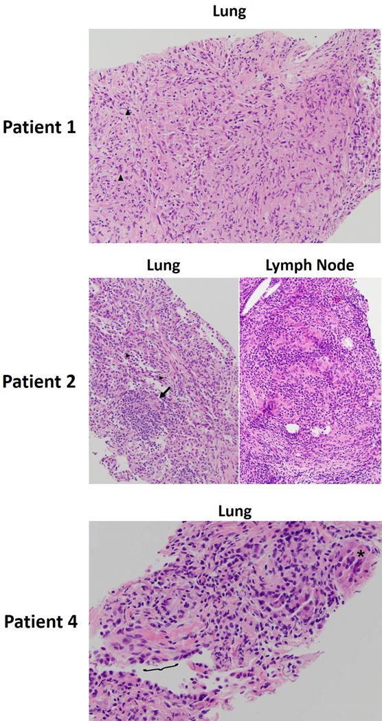
Figure 2. Histologic characteristics of sarcoid-like granulomatosis of the lung.

Patient 1: Hematoxylin and eosin (H&E) stain of the alveolated parenchyma of the lung. Inflammatory cells (predominantly polymorphonuclear cells) indicated by arrowheads. Patient 2: Left - H&E stain of the lung alveolar spaces. Eosinophils are indicated by arrowheads, and epithelioid giant cells indicated by the arrow. Right – H&E stain of the supraclavicular lymph node biopsy. Patient 4: core biopsy of the lung lesion. Peri-airway granulomas with prominent giant cells are indicated (asterisk). Terminal respiratory epithelium is denoted by the bracket.
