
Figure 2. MDA-9/Syntenin regulates PCa progression.
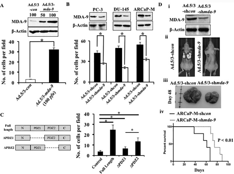
A) RWPE-1 cells infected with various adenoviruses at the indicated M.O.I. After 48 hours, infected cells were subjected to Matrigel invasion assays as described in Materials and methods. Expression of MDA-9/Syntenin was confirmed by Western blotting. B) MDA-9/Syntenin levels and invasiveness of different PCa cells after infection with Ad.5/3-shcon or Ad.5/3-shmda-9. C) Left panel: Schematic diagram of mutant mda-9/syntenin constructs, ΔPDZ1 and ΔPDZ2. Right panel: RWPE-1 cells transfected with full length and mutants of mda-9/syntenin and 48 hours after transfection invasion was evaluated. Number of invaded cells presented as a bar graph. Average with S.D. from three independent experiments. “*” represents statistical significance (p<0.05) between indicated groups. D) Downregulation of MDA-9/Syntenin was confirmed in Ad.shmda-9 infected cells (i). Luciferase expressing ARCaP-M cells carrying shcon or shmda-9 were inoculated I.V. into athymic nude mice by tail vein. Representative photographs of BLI (ii) and gross morphology of lungs (iii) 48 days after inoculation. Kaplan-Meier survival graph using Graph Pad software (iv).