
Figure 4. MDA-9/Syntenin plays a decisive role in IGFBP-2-induced STAT3 activation.
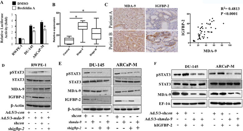
A) Cells transfected with a STAT3 reporter gene and after 36 hours were treated with Brefeldin A (5 µg/ml) for 30 min. Media was removed and cells were cultured an additional 3 hours in serum-free media and luciferase activity measured. B) Immunostaining for IGFBP-2 was performed in tissue microarray and staining intensity was quantified with Polaris Image Analysis software. The average value with S.D. from different stages is plotted. “*” represents statistical significance between groups. C) The expression of MDA-9/Syntenin and IGFBP-2 correlates in sections from the same patient (upper patient A (low expression) and patient B (high expression) bottom). Pearson correlation determined using 46 samples and presented with R2 and p value. D) RWPE-1 cells expressing MDA-9/Syntenin in presence of control shRNA or igfbp-2 shRNA for 48 hours. Cells were trypsinized and reseeded onto fibronectin-coated plates. Samples collected after 1 hour and Western blotting performed. E) Endogenous MDA-9/Syntenin and IGFBP-2 expression in PCa cells treated with shmda-9 and shigfbp-2 either alone or in combination. Cells were reseeded on fibronectin-coated plates for 1 hour. Western blotting with indicated antibodies. F) PCa cells infected with Ad.5/3-shcon or Ad.5/3-shmda-9 (100 pfu/cell) for 48 hours. Cells re-seeded on fibronectin-coated plates and treated with recombinant hIGFBP-2 (100 ng/ml) for 1 hour. Western blotting for indicated proteins.