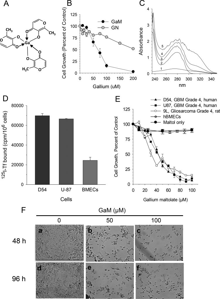
Figure 1.
GaM interaction with transferrin (Tf), Tf binding to cells, and effect of GaM on cell growth. A. Chemical structure of gallium maltolate. B. Comparison of the growth-inhibitory effects of gallium maltolate (GaM) and gallium nitrate (GN). D54 cells were incubated with equimolar concentrations of gallium as either GaM or GN. Cell proliferation was measured by MTT assay after 72-h of incubation. Values shown represent the means +/− S.E. (n = 3). C. GaM forms complexes with Tf in vitro. UV-visual spectra of 100 µM GaM (spectrum 1), 12.5 µM Tf (spectrum 2), and 12.5 µM Tf incubated with 12, 50, or 100 µM GaM (spectra 3, 4, and 5, respectively). Spectra were obtained at room temperature after a 30 minute incubation of Tf with GaM. D. TfR expression on glioblastoma cells and brain microvascular endothelial cells (BMECs). Specific 125I-Tf binding to TfR1 on intact cells is shown. E. GaM inhibits the growth of glioblastoma cell lines but not human brain microvascular endothelial cells (hBMECs). Maltol alone does not inhibit D54 cell growth. Cell growth was measured by MTT assay after a 5-day incubation of cells with GaM. Values shown represent means ± S.E. (n=3). F. Gallium maltolate induces cell death. Photomicrographs showing morphologic changes consistent with GaM-induced cell death in D54 cells incubated without or with GaM for 48 h (panels a, b and c) or 96 h (panels d, e, and f), respectively.

