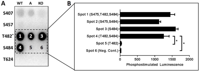
Figure 5.

(A) KCNQ1 carboxy peptides were exposed to activated δCaMKII. Each row contains peptides corresponding to the labeled KCNQ1 residue with columns for WT, A (phospho-acceptor site mutated to alanine), or KO (all serine and threonine mutated to alanine with the exception of T482 wherein S484 was not mutated; n=5 for each condition). (B) Quantification of phosphostimulated luminescence for WT, A, and KO peptides corresponding to KCNQ1 T482 and S484 during exposure to activated δCaMKII. *p<0.05, ns = not significant
