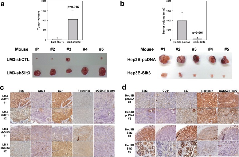
Fig. 4.
Slit3 negatively regulated HCC cell growth in vivo. a Stable Slit3 repressed clones (LM3-shSlit3) displayed induced tumor growth when compared with the control cells (LM-shCTL) 6 weeks post-injection of cells subcutaneously into flank region of nude mice (n = 5). b Stable Slit3 overexpressed clones (Hep3B-Slit3) displayed impaired tumor growth when compared with the control cells (Hep3B-pcDNA) 6 weeks post-injection of cells subcutaneously into flank region of nude mice (n = 5). c Immunohistochemical staining of Slit3, CD31, p27, β-catenin and phosopho-GSK3β (ser9) in tumor formed by LM3-shCTL and LM3-shSlit3 stable cells. d Immunohistochemical staining of Slit3, CD31, p27, β-catenin and phosopho-GSK3β (ser9) in tumor formed by Hep3B-pcDNA and Hep3B-Slit3 stable cells. Representative results from two mice of each group were shown

