Fig. 6.
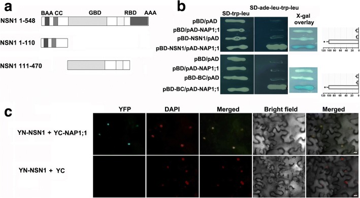
Interaction between NSN1 and NAP1;1 in yeast and tobacco. a Illustration of NSN1 fragments used for yeast two-hybrid assays. NSN1 protein consists of 548 amino acids. Two fragments tested in the yeast two-hybrid assay are the N-terminal fragment (amino acids 1–110) including the basic amino acid (BAA) domain and coiled-coil (CC) domain, and the fragment of amino acids 111–470 representing the GTP binding domain (GBD) and RNA binding domain (RBD). b Analysis of in vitro interaction between NAP1;1 and NSN1 with or without a domain deletion using the yeast two-hybrid system. The full length NSN1 was cloned into pBD vector as bait to screen a library of flower from ABRC (upper panel). A positive interaction colony was confirmed to contain AtNAP1;1 (At4g26110.2) by DNA sequencing. pAD-NAP1;1 represents the full length CDS of AtNAP1;1 cloned into pAD vector. The upper panel displays the interaction between AtNAP1;1 and NSN1; The lower panel shows the interactions between AtNAP1;1 and the N-terminal fragment of NSN1 including the basic amino acid (BAA) domain and coiled-coil (CC) domain, which was abbreviated as BC. Empty vectors pBD and pAD were used as negative controls. X-gal overlay assays and the quantification of ß-galactosidase activity were shown (right panels). Significant difference (p < 0.001) was evaluated by Student’s t test. c Analysis of in vivo interaction between NSN1 and NAP1;1 using biomolecule fluorescence complementation (BiFC) in tobacco leaves. An enhanced GFP (eGFP) was used [35] to test the interaction by fusing the coding sequence of NSN1 and AtNAP1;1 with the N-terminal (YFPN-NSN1) and the C-terminal fragment of GFP (YFPC-NAP1;1), respectively. The two constructs were co-expressed in tobacco leaves by Agrobacterium-infiltration. Infiltrated leaf epidermal cells were checked with confocal laser scanning. The co-expression of YFPN-NSN1 and no-fusion pSPYCE (M) 155 (YC) was used as a negative control. Nuclei were stained with DAPI. Scale bars: 10 μm

