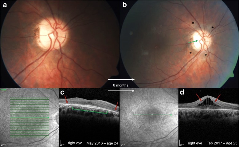
Fig. 2.
Tapeto-retinal degeneration in α-mannosidosis in two siblings. Fundus photographs (a, b) of the sister revealed progression of retinal pigment epithelium (RPE) atrophy outside the macula with yellow-white deposits around the optic disc and chorioretinal atrophy (black stars, b), and partial optic nerve atrophy (blue arrow, b). Optical coherence tomography (OCT) showed perifoveal thinning of the outer retinal layers and RPE (red arrows, c) with normal retinal layers in the fovea (green bracket, c). A progression of the outer retina thinning was seen and a cystic macula edema has developed within a year’s time at the age of 25 (red arrows, d)

