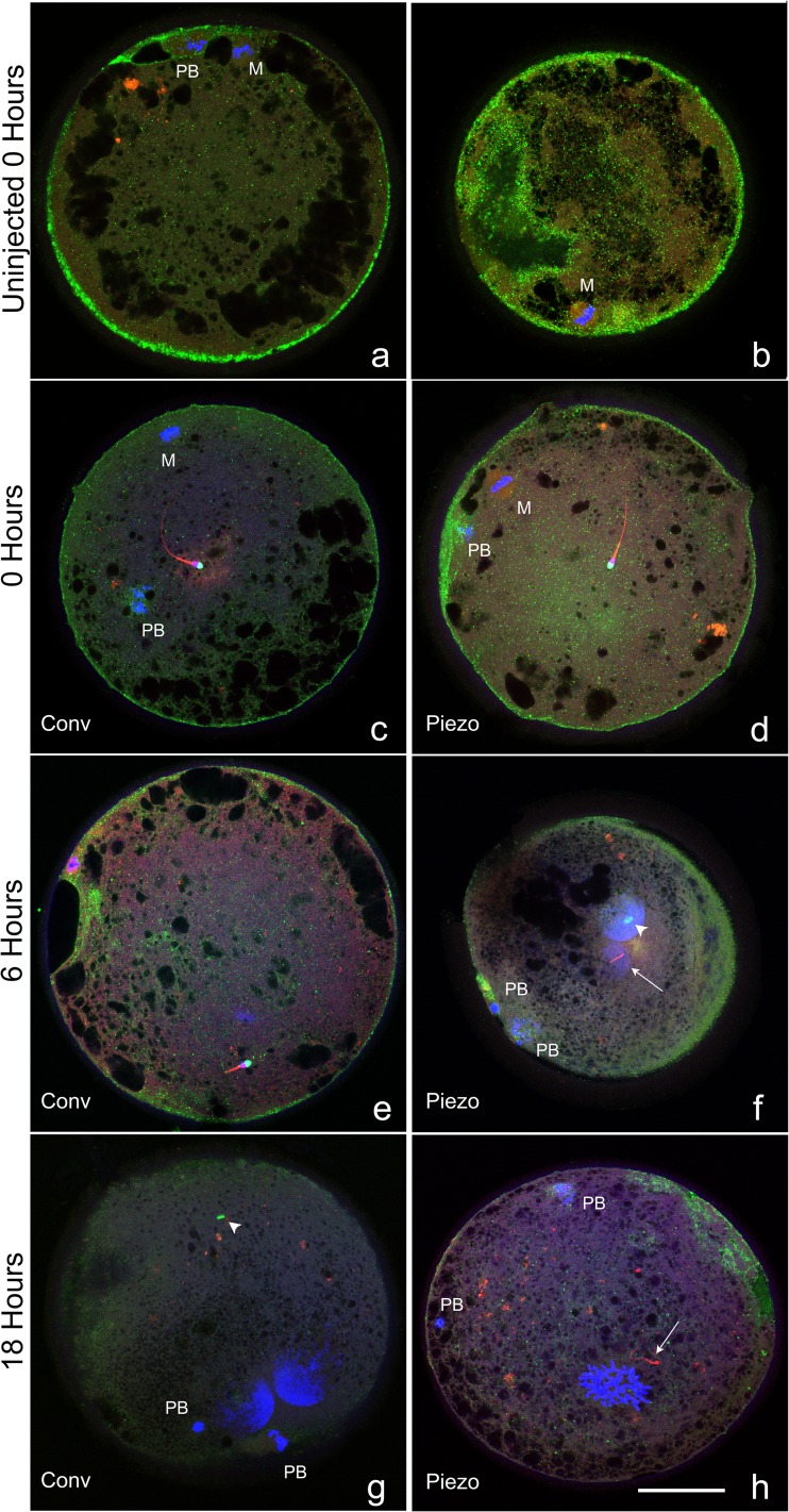
Fig. 2.
Confocal images of oocytes that were uninjected, or at 0, 6, or 18 h after conventional or Piezo ICSI, demonstrating different classifications of chromatin (DAPI, blue), acrosome (PNA, green), tail (MitoTracker Red CMXRos, red), and cortical granules (PNA, green). a–e MII oocytes (where visible; M oocyte metaphase plate, PB first polar body) each demonstrating a central sperm with intact sperm tail and acrosome. f, g Oocytes demonstrating two mid-stage pronuclei (large blue masses) and two polar bodies (PB), the acrosome is visible in the cytoplasm (arrowhead, green). The sperm tail is visible in (f) (arrow), and was present in the cytoplasm of (g) but is not visible in this focal plane. h Oocyte in prometaphase of the first mitotic division (blue area toward 6:00), with two polar bodies (PB). Sperm tail is visible in cytoplasm (arrow), no acrosome was visualized. Cortical granules were classified as prominent (a, b), moderate (c–e), and trace (f–h). Bar = 50 μm

