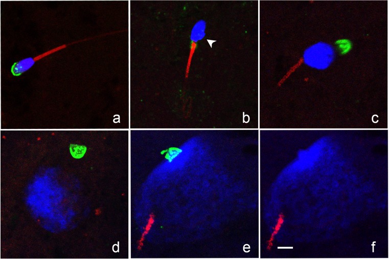
Fig. 4.
Confocal images of sperm chromatin (DAPI, blue), acrosome (PNA, green), and tail (MitoTracker Red CMXRos, red) showing different configurations. a Condensed sperm head with intact acrosome and tail. b Sperm head showing initial signs of decondensation (irregular protrusion of chromatin at base of head; arrowhead). Sperm tail is intact; acrosome was not visualized. We noted that remnants of the proximal droplet stained with PNA (green fluorescence on the midpiece at the junction of the sperm head and tail). c decondensing sperm chromatin, intact tail, and detached acrosome. d Early pronucleus with detached acrosome. e, f Mid-pronucleus with adjacent sperm tail; acrosome remained intact and chromatin within the acrosome failed to decondense, the green channel is removed in (f) to demonstrate chromatin clearly. Bar = 5 μm

