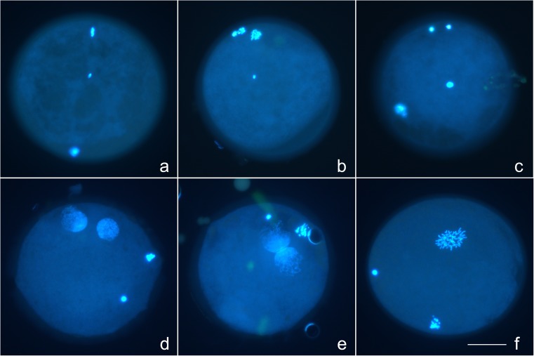
Fig. 5.
Photomicrographs taken on the inverted fluorescent microscope, demonstrating stages of oocyte activation and sperm head decondensation. a Metaphase II oocyte (metaphase plate at 12:00 and first polar body at 6:00) with central condensed sperm head. b Anaphase II with first polar body at 11:00, showing signs of division, and separating anaphase chromosomes at 12:00, with central condensed sperm head. c Telophase II with two masses of oocyte chromatin at 12:00 and first polar body at 8:00, with central decondensing sperm head. d Early pronuclear-stage oocyte with pronuclei at 12:00 and 1:00, and two polar bodies (3:00 and 4:00). e Late pronuclear-stage oocyte with pronuclei in contact with one another, and two polar bodies (12:00 and 2:00). f Oocyte in prometaphase of first mitosis, with two polar bodies (6:00 and 9:00). Note apparent cytoplasmic extrusion cone in perivitelline space between 2:00 and 3:00. Bar = 50 μm

