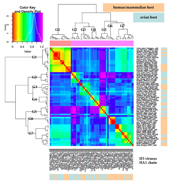
Figure 1.
ED analysis (heatmap) of a sixty-four haemagglutinin HA1 chains dataset from H5 viruses. As reported in the density plot, the warmer the colour, the lower the ED. Group numbers are established as explained in the main text; right and bottom bars highlight distribution of viruses isolated from either avian or human/mammalian host, according to indicated colour coding.

